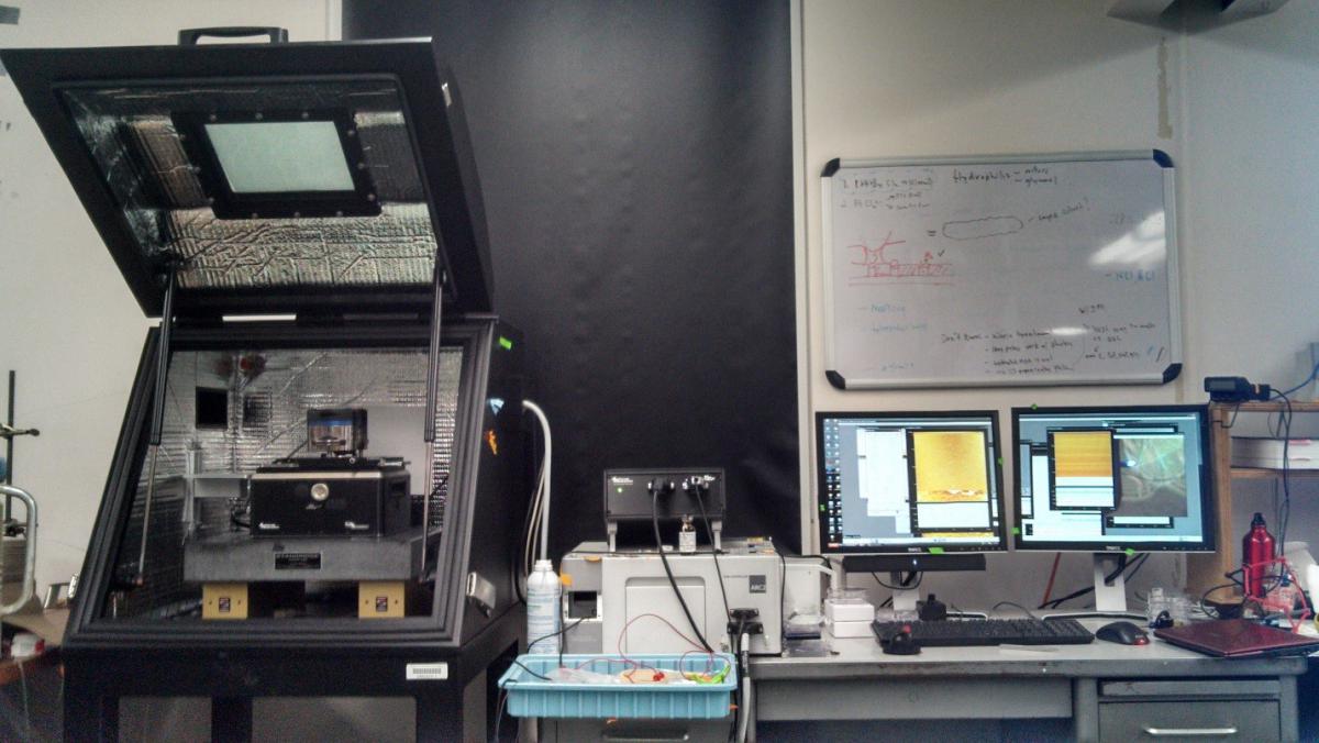
Asylum Research MFP-3D SA Atomic Force Microscope
This commercially available atomic force microscope in located in PSBN 4624E and allows for fast and reliable imaging with nanometer resolution. The MFP-3D (shown below) and corresponding software allow for a variety of imaging modes such as contact, AC mode, and FM mode and is useful for both soft and hard materials.
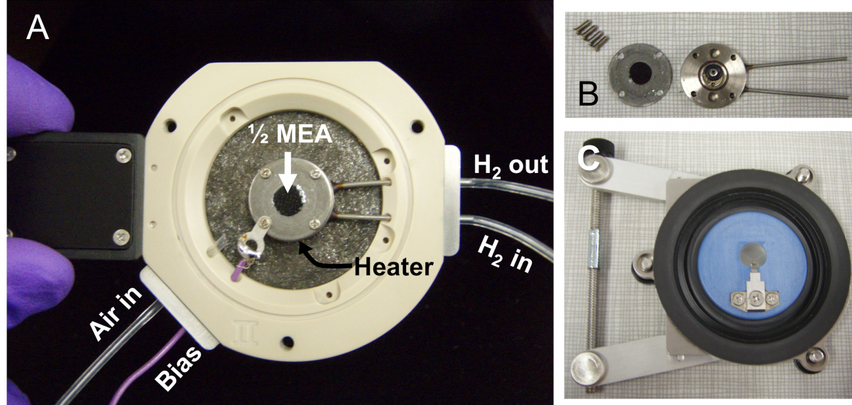
Using a closed fluid cell (A) with an internal heating stage we can precisely control the temperature and relative humidity of the sample which allows for the swelling and thermal rearrangement of polymeric samples to be probed. Our lab also possesses a conductive ORCA cantilever holder which allows for conductive imaging and electrical and electrochemical characterization of samples at the nanometer scale. Our specially designed nano fuel cell (B) can be placed inside of the closed fluid cell and allows for conductive AFM of ionomers such as Nafion® under fuel cell operating conditions.
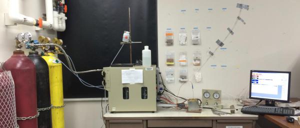
Scribner 850C compact Fuel Cell Test Station
This fuel cell test station allows us to characterize the electrochemical performance of fuel cells in house. This Station can operate at up to 100C, gas humidifiers and a back pressure module allow for the humidity, pressure and composition of reactant gases to be controlled precisely.
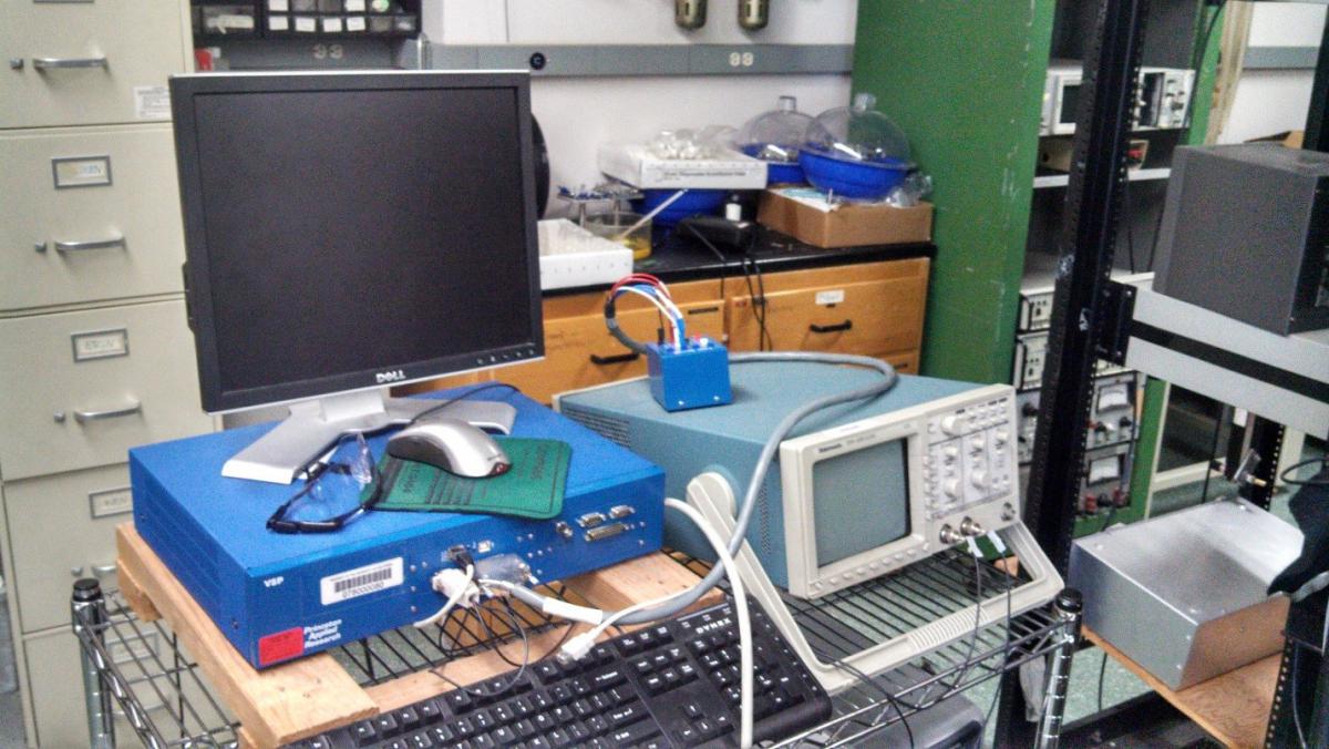
Princeton Applied Research (BioLogic) VSP model potentiostat
This potentiostat allows for a variety of electrochemical characterization techniques such as cyclic voltammetry and chronoamperometry with very high current and voltage resolution. In tandem with different electrodes and deposition cells it allows for electrochemical deposition of platinum and other metals onto catalytic supports of interest.
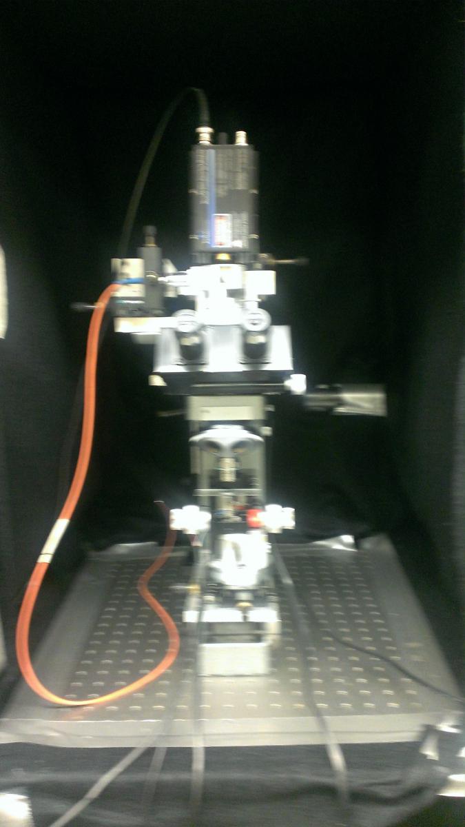
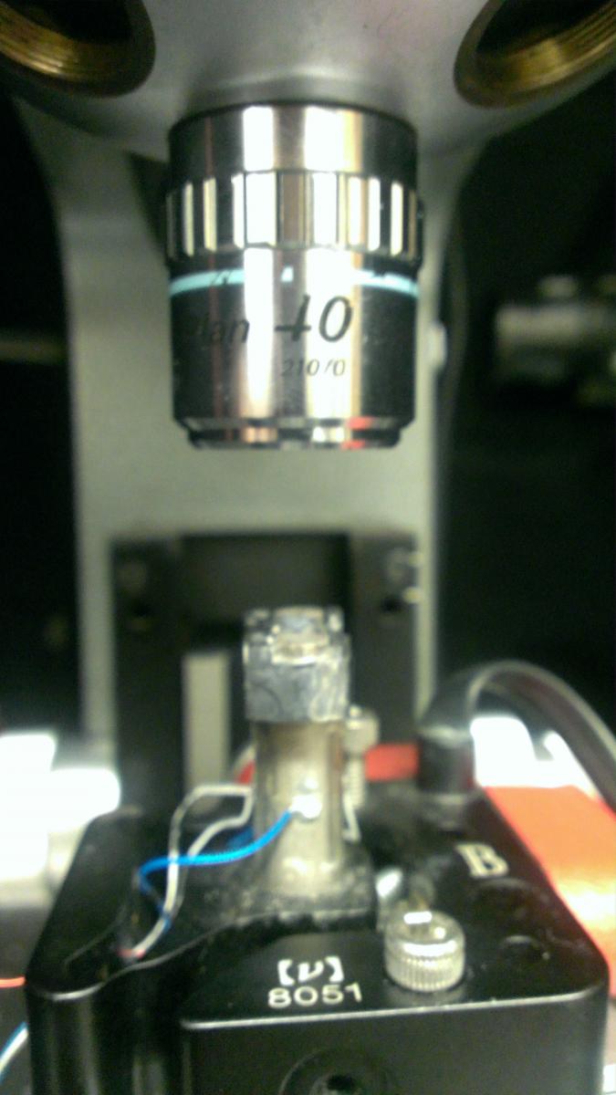
Laser Scanning Confocal Microscope
Laser Scanning Confocal Microscopy is a technique which utilizes a combination of pinhole optics and a high numerical aperture lens to focus a laser beam to a small area in order to achieve optical resolution which far exceeds that of traditional light microscopy. In our set-up the laser beam path is held stationary while the sample is raster scanned in order to obtain an image. Our home-built LSCM is used to analyze thin films as well as to do single molecule spectroscopy of conjugated oligomers. By swapping out for a variety of different scan heads, a range of scan sizes can be acheived. Furthermore, by utilizing a home built vacuum scan head, systems can be probed under a variety of ambient conditions. In the past, this instrumentation has also been applied by the Buratto group to probe a variety of systems including porous silicon, silver nanoparticles, and polyelectrolyte films.
